Discover 5 Unique Electron Microscopes from #brnoregion
Electron microscopy thrives in #brnoregion. Companies like Thermo Fisher Scientific, Delong Instruments, and Tescan have established themselves here, earning Brno global acclaim with their microscopes. From a small electron microscope that fits on a sturdy table to microscopes for observing frozen samples and live organisms—#brnoregion has it all. But what are these devices good for?
LVEM 5: The Smallest Electron Microscope in the World
“They are small, require minimal conditions, and even students can operate them. Our goal is to open the door to electron microscopy for everyone who needs it,” says Radka Martinková from Delong Instruments. This is where they developed this type of low-voltage electron microscope (LVEM), aiming to make electron microscopy accessible to a broader “audience.” The advantage is not only in the price. While it is in the range of a luxury car, LVEM devices from Delong are on the lower end of their category. Despite this, they are top-quality, which is why they are often among the first electron microscopes customers acquire.
The LVEM 5 from Delong Instruments weighs about seventy kilograms and can be placed on a sturdy table, making it the smallest electron microscope in the world. “The LVEM 5 allows for sample analysis in up to four different ways, which is why it is often acquired by teams needing multiple types of analysis but unable to afford a specialized device for each,” adds Martinková.
These microscopes are used in hospitals, research institutes, companies, and universities, helping detect viruses, bacteria, and other toxins that threaten the health of humans, animals, or plants and contaminate our environment.
MAMUT: Perfect Imaging of 2D Samples
The first device has nothing to do with the prehistoric animal, except perhaps its size. It is an electron microscope developed at the Institute of Scientific Instruments of the Czech Academy of Sciences. This advanced device operates in an ultra-high vacuum environment. This means the microscope contains a very low air pressure, minimizing electron scattering and improving the quality of the resulting image. Additionally, it uses very slow electrons, which have lower energy and allow for more detailed and precise information about surface structure and properties. MAMUT is useful wherever very clean surfaces need to be studied.
Eliška Materna Mikmeková from the mentioned institute, who leads the surface microscopy and spectrometry research team, uses it in her research on nanocrystals and their potential application in fuel production, in collaboration with colleagues from Rutgers University in the USA: “I use MAMUT primarily for studying so-called 2D materials, such as graphene, where I can observe these ‘invisible’ atom-thick samples with high resolution thanks to the very low energies. At very low energies, I can even determine if a sample is one or two atoms thick, showing atomic-level sensitivity! The ultra-high vacuum ensures that I can ‘observe,’ or scan, the sample with an electron beam without contaminating it with residual gases from the microscope chamber.”
TITAN with Atomic Resolution
An ideal companion for developing new materials and nanotechnologies in atomic scale is the state-of-the-art transmission electron microscope called TITAN. It is capable to operate with acceleration voltages from 60 kV up to 300 kV, allowing to study beam sensitive 2D samples as graphene, or, to achieve the highest image resolutionand electron transparency of thicker samples. This microscope is equipped with a monochromator ensuring low voltage imaging with substantially reduced chromatic aberrations and electron energy loss spectroscopy with high energy resolution. Its optical system also contains an image corrector limiting objective lens aberrations and providing TEM images with an ultimate atomic resolution deep below 1 angstrom. Above that, the microscope is equipped with a set of sensitive detectors for a variety of imaging and spectroscopy techniques.
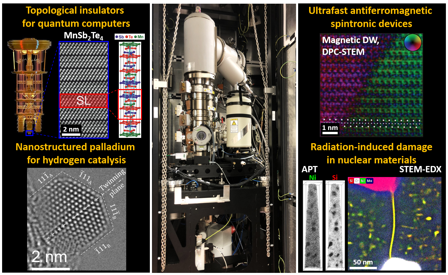
Simply put, TITAN can display observed samples with clearly distinguishable atoms and provide details about their chemical or physical state in atomic scale, which is crucial for research in material sciences and nanotechnology. Therefore, the CEITEC Nano research infrastructure, where the TITAN is installed, is exactly the place in #brnoregion where you can study and develop nanotechnology and advanced materials at the top scientific level.
Cryo-Electron Microscope for Virus Research
The TESCAN AMBER Cryo allows samples to be examined at temperatures as low as -160°C. The low temperature makes it possible to visualize biomolecules in their natural state. This preserves the internal structure of cells without the need for modification or degradation of samples. This is especially useful for studying lipid structures, which play a key role in many biological processes. Scientists can thus better understand what happens inside cells and what mechanisms affect their health.In general, cryo-electron microscopy plays an important role in studying viruses and enzymes, with far-reaching implications for drug and vaccine development. Another representative of cryo-electron microscopes from #brnoregion, the Thermo Scientific Krios Cryo-TEM, helped determine the structure of the SARS-CoV-2 spike protein, which was crucial in the development of the COVID-19 vaccine.
Observing Live Organisms
The final mention isn’t about the device itself but rather a unique microscopy method. The Environmental Electron Microscopy research group from the Institute of Scientific Instruments of the Czech Academy of Sciences was the first in the world to observe live rotifers under high gas pressure and at low temperatures. This even led to the discovery of two new rotifer species and a detailed description of several other species.
Read more about the topic
As Europe’s deep-tech landscape accelerates, Brno stands out as a city where science turns into startups. At this year’s Velvet Innovation Conference, which explores courage and resilience in innovation, emerging projects took the stage at the Startup Showcase. ACTOSens, Citymind, Lightly, EpiQ and Veriteus are those to watch. Curated by the innovation agency JIC, these startups combine academic excellence with global ambition, addressing challenges from healthcare and AI to cybersecurity and industrial automation.